

Summary: A new AI tool, MindGlide, can rapidly analyze routine brain MRI scans to detect subtle changes caused by multiple sclerosis (MS), such as brain shrinkage and lesions. Traditionally requiring expert interpretation, this process now takes just seconds, allowing more efficient monitoring of disease progression and treatment effects.
In tests with over 14,000 images, MindGlide outperformed existing tools and proved reliable across different brain regions and scan types. Researchers hope it will soon unlock insights from millions of archived hospital scans and transform MS care.
Key Facts:
- Rapid Results: MindGlide analyzes brain MRIs in 5–10 seconds per image.
- High Accuracy: Outperformed two existing AI tools in detecting MS lesions.
- Clinical Potential: Can interpret routine scans previously unusable for MS analysis.
Source: UCL
A new artificial intelligence (AI) tool that can help interpret and assess how well treatments are working for patients with multiple sclerosis (MS) has been developed by UCL researchers.
AI uses mathematical models to train computers using massive amounts of data to learn and solve problems in ways that can seem human, including to perform complex tasks like image recognition.
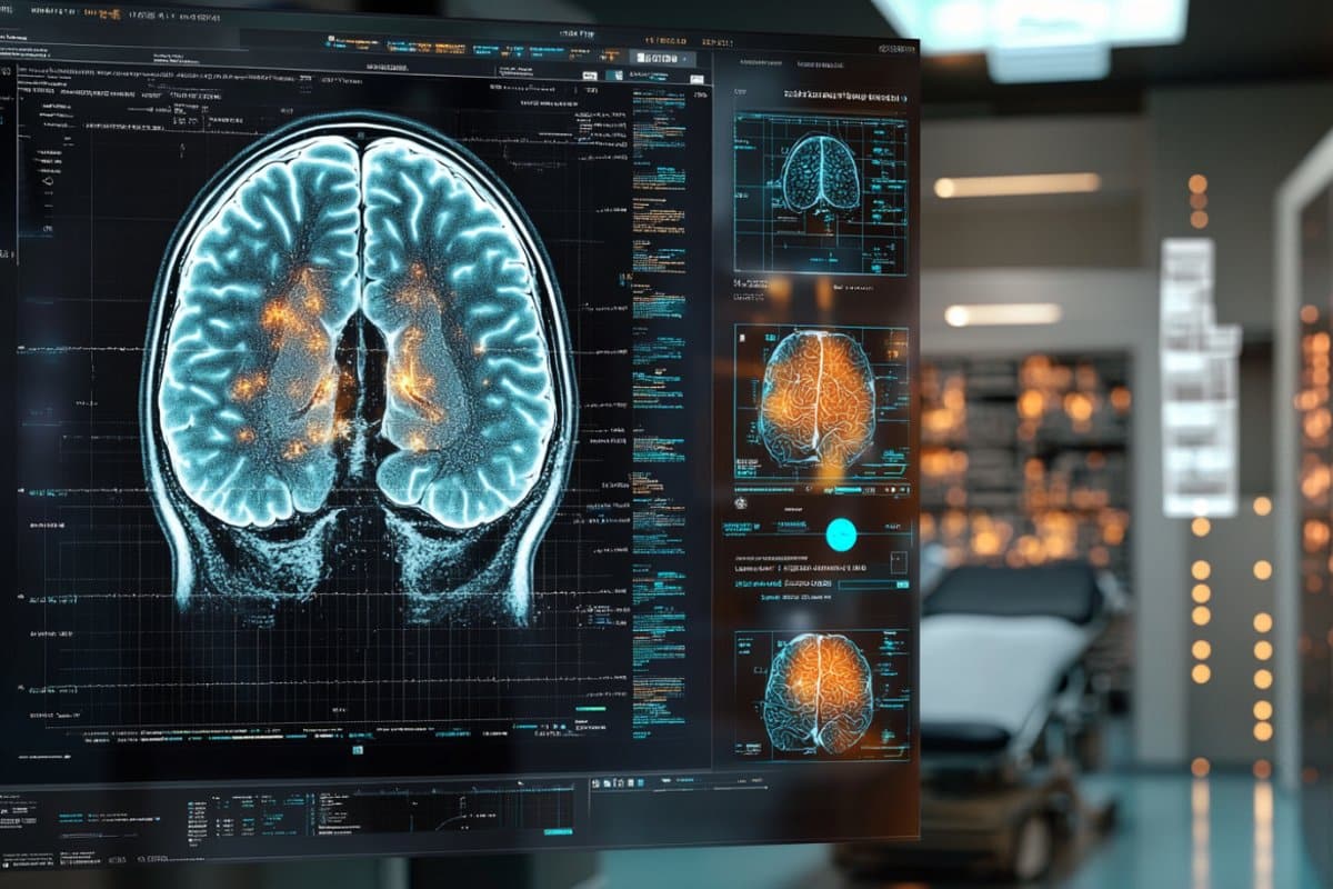
The tool, called MindGlide, can extract key information from brain images (MRI scans) acquired during the care of MS patients, such as measuring damaged areas of the brain and highlighting subtle changes such as brain shrinkage and plaques.
MS is a condition where the immune system attacks the brain and spinal cord. This causes problems in how a person moves, feels or thinks. In the UK, 130,000 people live with MS, costing the NHS more than £2.9 billion a year.
Magnetic Resonance Imaging (MRI) markers are crucial for studying and testing treatments for MS. However, measuring these markers needs different types of specialised MRI scans, limiting the effectiveness of many routine hospital scans.
As part of a new study, published in Nature Communications, researchers tested the effectiveness of MindGlide on over 14,000 images from more than 1,000 patients with MS.
This task had previously required expert neuro-radiologists to interpret years of complex scans manually – and the turnaround time for reporting these images is often weeks due to the NHS workload.
However, for the first time, MindGlide was able to successfully use AI to detect how different treatments affected disease progression in clinical trials and routine care, using images that could not previously be analysed and routine MRI scan images. The process took just five to 10 seconds per image.
MindGlide also performed better than two other AI tools – SAMSEG (a tool used to identify and outline different parts of the brain in MRI scans) and WMH-SynthSeg (a tool that detects and measures bright spots seen on certain MRI scans, that can be important for diagnosing and monitoring conditions like MS) – when compared to expert clinical analysis.
MindGlide was 60% better than SAMSEG and 20% better than WMH-SynthSeg for locating brain abnormalities known as plaques (or lesions) or for monitoring treatment effect.
First author, Dr Philipp Goebl (UCL Queen Square Institute of Neurology and UCL Hawkes Institute), said: “Using MindGlide will enable us to use existing brain images in hospital archives to better understand multiple sclerosis and how treatment affects the brain.
“We hope that the tool will unlock valuable information from millions of untapped brain images that were previously difficult or impossible to understand, immediately leading to valuable insights into multiple sclerosis for researchers and, in the near future, to better understand a patient’s condition through AI in the clinic. We hope this will be possible in the next five to 10 years.”
The results from the study show that it is possible to use MindGlide to accurately identify and measure important brain tissues and lesions even with limited MRI data and single types of scans that aren’t usually used for this purpose – such as T2-weighted MRI without FLAIR (a type of scan that highlights fluids in the body but still contains bright signals – making it harder to see plaques).
As well as performing better at detecting changes in the brain’s outer layer, MindGlide also performed well in deeper brain areas.
The findings were valid and reliable both at one point in time and over longer periods (i.e. at annual scans attended by patients).
Additionally, MindGlide was able to corroborate previous high-quality research regarding which treatments were most effective.
The researchers now hope that MindGlide can be used to evaluate MS treatments in real-world settings, overcoming previous limitations of relying solely on high-quality clinical trial data, which often did not capture the full diversity of people with MS.
Dr Arman Eshaghi (UCL Queen Square Institute of Neurology and UCL Hawkes Institute), the project’s principal investigator and lead of the MS-PINPOINT group, said: “We were not previously analysing the bulk of clinical brain images due to their lower quality. AI will unlock the untapped potential of the treasure trove of hospital information to provide unprecedented insights into MS progression and how treatments work and affect the brain.”
Study limitations
The current implementation of MindGlide is limited to brain scans and does not include spinal cord imaging, which is important for assessing disability in people with MS. Future research will need to develop a more comprehensive assessment of the whole neural system to encompass both the brain and the spinal cord.
Developing MindGlide
MindGlide is a deep learning (AI) model, developed by UCL researchers, to assess MRI images of the brain and identify damage and changes caused by MS.
In developing MindGlide scientists used an initial dataset of 4,247 brain MRI scans from 2,934 MS patients across 592 MRI scanners. During this process MindGlide trains itself to identify markers of the disease.
This new study was carried to validate MindGlide, against three separate databases of 14,952 images from 1,001 patients.
About this AI and MS research news
Author: Poppy Tombs
Source: UCL
Contact: Poppy Tombs – UCL
Image: The image is credited to Neuroscience News
Original Research: Open access.
“Repurposing Clinical MRI Archives for Multiple Sclerosis Research with a Flexible, Single-Contrast Approach: New Insights from Old Scans” by Philipp Goebl et al. Nature Communications
Abstract
Repurposing Clinical MRI Archives for Multiple Sclerosis Research with a Flexible, Single-Contrast Approach: New Insights from Old Scans
Magnetic resonance imaging (MRI) biomarkers are vital for multiple sclerosis (MS) clinical research and trials but quantifying them requires multi-contrast protocols and limits the use of abundant single-contrast hospital archives.
We developed MindGlide, a deep learning model to extract brain region and white matter lesion volumes from any single MRI contrast.
We trained MindGlide on 4247 brain MRI scans from 2934 MS patients across 592 scanners, and externally validated it using 14,952 scans from 1,001 patients in two clinical trials (primary-progressive MS and secondary-progressive MS trials) and a routine-care MS dataset.
The model outperformed two state-of-the-art models when tested against expert-labelled lesion volumes.
In clinical trials, MindGlide detected treatment effects on T2-lesion accrual and cortical and deep grey matter volume loss. In routine-care data, T2-lesion volume increased with moderate-efficacy treatment but remained stable with high-efficacy treatment.
MindGlide uniquely enables quantitative analysis of archival single-contrast MRIs, unlocking insights from untapped hospital datasets.
